Radioactive decay isn’t just something you hear about in science textbooks; it’s actually a quiet hero in the world of medicine, playing a surprisingly big role in how we diagnose and treat a variety of conditions. Forget the sci-fi movie depictions of glowing green mutants – in the medical realm, controlled radioactive materials, often called radioisotopes, are carefully used for some incredibly helpful purposes. So, in a nutshell, radioactive decay impacts medicine by allowing us to “see” inside the body without invasive surgery and to target and destroy diseased cells.
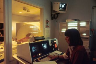
One of the most significant ways radioactive decay impacts medicine is through its use in diagnostic imaging. This technology allows doctors to get a detailed look at what’s happening inside your body, helping them identify problems that would otherwise be hidden. It’s a far cry from the days of simply looking at X-rays.
The Magic of Tracers
The fundamental principle behind these imaging techniques is the use of radioactive tracers. These are small amounts of radioactive substances that are introduced into the body, usually injected or swallowed. As these tracers go through your system, they emit radiation that is detected by special cameras. The decay process of the radioisotope is crucial here. The rate at which it decays (its half-life) is carefully chosen to be long enough for the imaging procedure but short enough to minimize the patient’s exposure to radiation.
PET Scans: Mapping Your Body’s Activity
Positron Emission Tomography (PET) scans are a prime example of this. In a PET scan, a radioactive tracer, often a form of glucose tagged with a positron-emitting isotope like Fluorine-18, is given to the patient. Cancer cells, for instance, often consume glucose at a higher rate than normal cells. As the tracer decays, it emits positrons, which collide with electrons in the body, producing gamma rays. These gamma rays are then detected by the PET scanner. The concentration of the tracer in different tissues reveals areas of high metabolic activity, which can pinpoint tumors, assess their spread, and see how well cancer treatment is working. It’s like giving your body a map of its own energy usage, highlighting where things might be going wrong.
SPECT Scans: A Different Perspective
Single-Photon Emission Computed Tomography (SPECT) uses a similar concept but with different types of radioactive tracers that emit gamma rays directly. These tracers can be designed to accumulate in specific organs or tissues. For example, radioisotopes of Technetium or Iodine are often used. SPECT scans are particularly useful for evaluating blood flow to organs like the heart and brain, identifying areas of infection or inflammation, and assessing bone health. The “tomography” in SPECT means it creates cross-sectional images, much like a CT scan, giving a 3D view of the distribution of the tracer. This can be invaluable for understanding brain activity in cases of epilepsy or stroke, or for detecting bone fractures that might not be visible on a standard X-ray.
Nuclear Medicine Scans: Broad Applications
Beyond PET and SPECT, a whole range of nuclear medicine scans utilize radioactive decay. Thyroid scans, for example, use radioactive iodine to check the function of the thyroid gland. This is vital for diagnosing conditions like hyperthyroidism or hypothyroidism. Bone scans can detect cancer that has spread to the bones, or identify areas of bone injury or infection. Kidney scans can assess the function of the kidneys. The ability to track the movement and accumulation of these radioactive substances provides a wealth of functional information about organs that traditional imaging techniques cannot offer.
Radioactive decay plays a crucial role in the field of medicine, particularly in diagnostic imaging and cancer treatment. For a deeper understanding of how these processes are utilized in medical applications, you can explore the article on the impact of radioactive isotopes in healthcare. This article provides insights into how radioactive decay is harnessed for both therapeutic and diagnostic purposes, enhancing our ability to detect and treat various medical conditions. To read more, visit this article.
Fighting Back: Radiation Therapy for Cancer
Another monumental impact of radioactive decay on medicine is in the treatment of cancer. Radiation therapy, also known as radiotherapy, harnesses the energy released from radioactive decay to destroy cancerous cells. This is a powerful tool that has saved countless lives.
External Beam Radiation Therapy: Precision Targeting
In external beam radiation therapy, a machine outside the body delivers radiation to the tumor. While the machine itself doesn’t contain a radioactive source that decays in the traditional sense (it often uses linear accelerators to generate high-energy X-rays), the principle of using energetic particles to damage DNA is directly linked to the understanding of radioactivity derived from studying natural decay processes. This allows for highly precise targeting of tumors while sparing surrounding healthy tissues. The energy and duration of the radiation are meticulously controlled to maximize cell destruction within the tumor and minimize side effects.
Internal Radiation Therapy (Brachytherapy): Delivering a Punch Up Close
Brachytherapy takes a more direct approach. Here, small radioactive sources (often called seeds, ribbons, or capsules) are placed directly inside or very close to the tumor. These sources decay over time, releasing radiation that targets and kills the cancer cells. This method is particularly effective for certain types of cancer like prostate cancer, cervical cancer, and breast cancer. The half-life of the isotopes used in brachytherapy is carefully selected. For instance, Iodine-125 has a half-life of about 59 days and is often used for permanent implants, while Iridium-192 has a shorter half-life of about 74 days and is used for temporary implants, allowing the radiation dose to be delivered over a specific period. This controlled decay is key to delivering the therapeutic dose effectively.
Targeted Radionuclide Therapy: A Smarter Attack
More advanced forms of radiation therapy, like targeted radionuclide therapy, use radioactive isotopes that are specifically attracted to cancer cells. These isotopes can be attached to molecules that bind to proteins on the surface of cancer cells. When the radioactive isotope decays, it delivers a high dose of radiation directly to the cancer cells, minimizing damage to healthy tissues. For example, Lutetium-177-DOTATATE is used to treat certain types of neuroendocrine tumors. The molecule DOTATATE binds to somatostatin receptors found on these tumors, delivering the radioactive Lutetium-177 directly to the cancerous cells. This is a brilliant application where the principles of radioactive decay are combined with molecular targeting for incredibly precise cancer treatment.
Beyond Imaging and Treatment: Other Medical Marvels
The impact of radioactive decay extends beyond these primary applications, touching various other aspects of medical care and research.
Sterilization: Keeping Medical Equipment Safe
One of the less “glamorous” but critically important uses of radioactive decay is in the sterilization of medical equipment. Gamma irradiation, often using Cobalt-60, is a highly effective method for killing bacteria, viruses, and other microorganisms on heat-sensitive medical devices like syringes, gloves, and surgical instruments. The gamma rays penetrate packaging and sterilize the products without damaging them. This process is efficient, reliable, and ensures that medical supplies are safe for use, which is fundamental to preventing infections and ensuring patient safety. The consistent and predictable decay of Cobalt-60 allows for precise control over the sterilization process.
Medical Research: Unraveling Biological Mysteries
Radioactive isotopes are also indispensable tools in medical research. By tagging molecules with radioisotopes, scientists can track the movement and fate of these molecules within living organisms. This allows researchers to study metabolic pathways, understand how drugs are absorbed and eliminated, and investigate the mechanisms of diseases. For example, researchers might use a radioactively labeled compound to study how a particular nutrient is processed in the body or to trace the spread of a disease at a cellular level. This fundamental understanding, driven by the ability to detect minute amounts of radioactive decay, is what leads to new diagnostic techniques and treatment strategies.
Dating and Forensics: A Surprisingly Relevant Connection
While not directly part of patient care, the principles of radioactive decay are also foundational to techniques that have indirect medical relevance. Radiocarbon dating, for instance, allows us to understand the age of ancient biological samples, which can inform our understanding of the evolution of diseases and pathogens. In forensic medicine, isotopic analysis can help determine the origin of drugs or biological samples, aiding in investigations. These applications, while not clinical treatments, highlight the pervasive importance of understanding radioactive decay.
The Science Behind the Rays: Understanding Half-Life and Isotopes
To truly appreciate how radioactive decay impacts medicine, it’s helpful to understand a couple of key concepts: isotopes and half-life. They are the bedrock of why these medical applications are possible and safe.
Isotopes: Different Faces of the Same Element
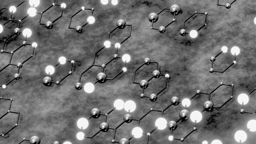
Every element on the periodic table is defined by the number of protons in its nucleus. However, atoms of the same element can have different numbers of neutrons. These variations are called isotopes. Some isotopes are stable, meaning their nucleus doesn’t change spontaneously. Others are unstable, or radioactive, and their nuclei will undergo radioactive decay to become more stable. In medicine, we specifically use these radioactive isotopes, also known as radioisotopes or radionuclides.
Half-Life: The Predictable Clock of Decay
The “half-life” is a fundamental property of a radioactive isotope. It’s the time it takes for half of the radioactive atoms in a sample to decay. Each radioisotope has a unique, predictable half-life, ranging from fractions of a second to billions of years. This predictability is crucial for medical applications.
For diagnostic imaging, radioisotopes with short half-lives are often preferred. This means they decay quickly, delivering their radiation dose to the patient and then becoming inactive within a reasonable timeframe, minimizing long-term radiation exposure. For example, Technetium-99m, a workhorse in nuclear medicine, has a half-life of about six hours, making it ideal for scans that need to be completed within a day.
For radiation therapy, the choice of isotope and its half-life depends on the treatment strategy. Some therapies use isotopes with longer half-lives to deliver a sustained dose over weeks or months, as in some forms of brachytherapy. Others might use isotopes with shorter half-lives for a more intense, localized dose over a shorter period. The selection is always a careful balance of efficacy and safety.
Radioactive decay plays a crucial role in the field of medicine, particularly in diagnostic imaging and cancer treatment. The process allows for the use of radioactive isotopes in various medical applications, such as PET scans and targeted radiotherapy, which help in detecting diseases and treating tumors effectively. For a deeper understanding of how these principles are applied in modern medicine, you can read more in this insightful article about the advancements in medical technology and their implications for patient care. Check it out here.
Safety and Regulation: Ensuring Responsible Use
| Aspect | Impact |
|---|---|
| Radioactive isotopes | Used in medical imaging to diagnose and treat diseases |
| Radiation therapy | Utilizes controlled radioactive decay to target and destroy cancer cells |
| Radioisotope therapy | Administered to treat conditions such as hyperthyroidism and certain types of cancer |
| Radioactive tracers | Used to track the movement of substances within the body for diagnostic purposes |
The use of radioactive materials in medicine isn’t taken lightly. Strict regulations and protocols are in place to ensure the safety of patients, healthcare professionals, and the public.
Minimizing Exposure: The ALARA Principle
A core principle guiding the use of radiation in medicine is “As Low As Reasonably Achievable” (ALARA). This means that radiation doses are kept as low as possible while still achieving the diagnostic or therapeutic goal. This involves careful selection of radioisotopes, precise calibration of equipment, and efficient imaging or treatment techniques.
Shielding and Containment: Protecting Against Radiation
Radioactive materials are handled with extreme care. Lead shielding is commonly used to block radiation. Radioactive waste is managed and disposed of according to strict guidelines to prevent environmental contamination. Healthcare professionals who work with radioactive materials undergo specialized training to ensure they understand and follow safety procedures.
Regulatory Oversight: Keeping Everyone Accountable
Government agencies and international bodies set standards and regulations for the use of radioactive materials in medicine. These regulations cover everything from the licensing of facilities to the training of personnel and the safe disposal of radioactive waste. This oversight ensures that the powerful tools of radioactive decay are used responsibly and ethically for the benefit of patients.
In conclusion, the impact of radioactive decay on medicine is profound and multifaceted. From offering unprecedented views inside the human body, to directly combatting diseases like cancer, to ensuring the sterility of medical supplies, this natural phenomenon, when harnessed with scientific rigor and ethical consideration, has become an indispensable pillar of modern healthcare. It’s a testament to human ingenuity that we can take something as powerful and potentially dangerous as radioactive decay and transform it into a force for healing and discovery.

Why Modern Medicine Can’t Store Tomorrow
FAQs
What is radioactive decay?
Radioactive decay is the process by which an unstable atomic nucleus loses energy by emitting radiation. This process can result in the transformation of the original atom into a different element.
How does radioactive decay affect medicine?
Radioactive decay is used in medicine for various purposes, including medical imaging, cancer treatment, and sterilization of medical equipment. For example, radioactive isotopes can be used as tracers to diagnose and monitor diseases, and radiation therapy can be used to target and destroy cancer cells.
What are some examples of radioactive isotopes used in medicine?
Some examples of radioactive isotopes used in medicine include technetium-99m for medical imaging, iodine-131 for thyroid cancer treatment, and cobalt-60 for radiation therapy. These isotopes emit radiation that can be used for diagnostic and therapeutic purposes.
What are the risks associated with using radioactive decay in medicine?
While radioactive decay has many benefits in medicine, there are also risks associated with exposure to radiation. These risks include potential damage to healthy cells, radiation sickness, and long-term effects such as an increased risk of cancer. It is important for medical professionals to carefully weigh the benefits and risks when using radioactive decay in medicine.
How is radioactive decay regulated in medicine?
The use of radioactive materials in medicine is regulated by government agencies such as the Nuclear Regulatory Commission (NRC) in the United States. These agencies set standards for the safe use and handling of radioactive materials, as well as for the training and certification of personnel working with these materials. Strict regulations and guidelines are in place to ensure the safe and effective use of radioactive decay in medicine.