So, you’re curious about “Unveiling the Power of Nuclear Medicine: A Science Documentary,” and what this film is all about, right? Well, the short answer is: it’s a deep dive into how we use tiny amounts of radioactivity, not to destroy, but to heal and diagnose within the human body. Think of it as using a very sophisticated, internal X-ray that shows us what’s happening at a cellular level, and in many cases, how we can target and treat diseases like cancer more precisely than ever before. This documentary aims to demystify this complex field, making it accessible and highlighting its incredible impact on modern healthcare.
Before we get into the nitty-gritty of treatments and scans, it’s crucial to get a handle on what makes nuclear medicine tick. At its core, it’s all about radioisotopes. These are essentially atoms of an element that have an unstable nucleus, and because of that instability, they release energy in the form of radiation. Now, that word, “radiation,” can sound a bit scary, but in nuclear medicine, we’re talking about carefully controlled, incredibly small doses.
What Exactly Are Radioisotopes?
Think of atoms as tiny building blocks. Most atoms are perfectly happy and stable. Radioisotopes are like those same building blocks, but with just a little bit of extra “oomph” inside their core. This extra “oomph” makes them unstable, and they naturally tend to shed this instability by emitting particles or energy. This emission is what we call radiation.
Not All Radiation is Created Equal
It’s important to understand that not all radiation is the same. Nuclear medicine primarily uses two types: alpha and beta particles, and gamma rays. Alpha and beta particles are relatively low-energy and don’t travel far. They’re often used in targeted therapies, staying close to where they’re administered. Gamma rays, on the other hand, are higher energy and can travel further, which is why they’re perfect for imaging – they escape the body and can be detected by special cameras. The key is that the radioisotopes we use are chosen for their specific properties, ensuring they emit the right kind of radiation for the job, and importantly, that they don’t stick around in the body for too long.
Where Do These Isotopes Come From?
The “ingredients” for nuclear medicine are produced in a few key ways. Some radioisotopes are created in nuclear reactors, where specific materials are bombarded with neutrons. Others are generated in cyclotrons, which are particle accelerators that smash protons into target atoms. For some of the shorter-lived isotopes, they’re actually produced on-site at hospitals using a device called a generator, which “milks” a parent radioisotope to get a short-lived daughter isotope. This is a fascinating logistical dance, ensuring that these very specific, often short-lived, medical tools are available when and where they’re needed.
Nuclear medicine is a fascinating field that combines advanced imaging techniques with therapeutic applications, making it a crucial component of modern healthcare. For those interested in exploring this topic further, a related article can be found at this link, which delves into the latest advancements and the impact of nuclear medicine on patient care. This documentary-style piece provides insights into how radioactive materials are used in diagnosis and treatment, highlighting the importance of this innovative branch of medicine.
Seeing the Unseen: Diagnostic Imaging in Nuclear Medicine
One of the most visually striking aspects of nuclear medicine is its ability to create images that show us the body functioning at a molecular level. This goes far beyond what traditional X-rays or CT scans can offer, which are excellent at showing structure but less adept at revealing biological processes.
The Magic of Radiopharmaceuticals
The stars of diagnostic nuclear medicine are radiopharmaceuticals. These are compounds that have been “tagged” with a radioisotope. The trick is that these compounds are designed to naturally go to specific organs, tissues, or even processes within the body. For example, a radiopharmaceutical might be designed to be taken up by bone cells, or by certain types of tumors, or by the thyroid gland.
Bone Scans: Uncovering Hidden Fractures
Consider a bone scan. If you suspect a stress fracture that might not show up on a regular X-ray, a bone scan can be incredibly useful. The radiopharmaceutical is injected into the bloodstream and travels throughout the body. It’s preferentially taken up by areas of increased bone turnover, which often indicates where there’s been damage or healing. The gamma rays emitted by the radioisotope are then detected by a gamma camera, creating an image that highlights these areas of high activity, pinpointing the fracture with remarkable accuracy.
PET Scans: Mapping Metabolic Activity
Positron Emission Tomography (PET) scans are another powerful diagnostic tool. PET scans use radiopharmaceuticals that emit positrons. When a positron encounters an electron in the body, they annihilate each other, producing two gamma rays that travel in opposite directions. The PET scanner detects these pairs of gamma rays, allowing for incredibly detailed 3D images that show metabolic activity. This is particularly useful in oncology, where doctors can see if a tumor is actively growing and spreading by looking at its glucose metabolism.
Beyond Bones and Tumors: A Spectrum of Applications
The diagnostic applications are incredibly broad. We have scans to assess heart function, evaluate brain activity in conditions like Alzheimer’s or epilepsy, check kidney function, and diagnose thyroid disorders, among many others. The ability to see how organs are working, not just what they look like, is a game-changer for early detection and precise diagnosis.
Targeted Therapies: Radioisotopes as Tiny Warriors
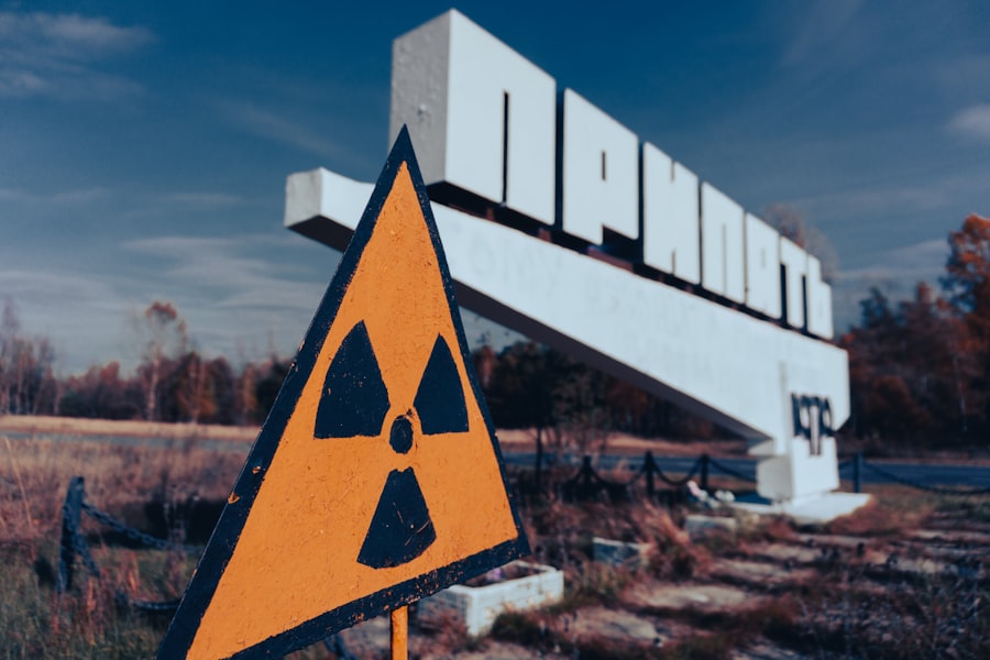
While diagnostics are crucial, the power of nuclear medicine really shines in its therapeutic applications. Here, radioisotopes are used not to see, but to treat. The concept is elegant: deliver a radioactive substance directly to diseased cells, where its radiation can effectively destroy them while minimizing damage to surrounding healthy tissue.
The Art of Precision Targeting
The success of radionuclide therapy hinges on delivering the radioisotope specifically to where it’s needed. This is often achieved by attaching the radioisotope to a molecule that has a high affinity for cancer cells or other diseased tissues. This is where the documentary likely delves into specific examples.
Iodine-131 for Thyroid Cancer
A classic example is the use of Iodine-131 to treat certain types of thyroid cancer. The thyroid gland naturally absorbs iodine. When a patient with thyroid cancer ingestable radioactive iodine, the thyroid cells, including the cancer cells, take it up. The radiation then targets and destroys these cells. This is a highly effective treatment that has saved countless lives.
Lutetium-177 PSMA Therapy for Prostate Cancer
More recently, therapies like Lutetium-177 PSMA have revolutionized the treatment of advanced prostate cancer. PSMA (prostate-specific membrane antigen) is a protein that is often overexpressed on prostate cancer cells. A molecule that binds to PSMA is linked to Lutetium-177, a therapeutic radioisotope. When injected, this complex seeks out and binds to prostate cancer cells, delivering its radiation directly to the tumor cells. This offers a highly targeted and often well-tolerated treatment option for patients with metastatic disease.
The Future is Radioligand Therapy
Radioligand therapy is a rapidly evolving field. The principle is to use a specific ligand (the molecule that targets the disease) attached to a therapeutic radioisotope. This allows for incredibly precise delivery of radiation, reducing side effects and improving patient outcomes. The documentary might explore ongoing research and clinical trials exploring this powerful approach for a widening range of cancers and other diseases.
The Science Behind the Scans: Equipment and Techniques
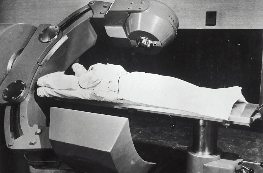
Seeing these incredible images and delivering targeted therapies requires sophisticated equipment and highly specialized techniques. The documentary likely dedicates time to explaining how these seemingly magical processes actually work on a technical level.
Gamma Cameras: The Eyes of Nuclear Medicine
The workhorse of many nuclear medicine imaging procedures is the gamma camera. After a radiopharmaceutical is administered, it emits gamma rays as it concentrates in specific areas of the body. The gamma camera, with its many crystal detectors, captures these gamma rays. The timing and location of the detection are processed by computer systems to create detailed, often 3D, images that show how the radiopharmaceutical has been distributed.
PET Scanners: Unraveling Metabolic Maps
PET scanners are even more advanced. They are designed to detect the pairs of gamma rays produced by positron annihilation. This requires a ring of detectors surrounding the patient, capable of capturing these signals from multiple angles simultaneously. The sophisticated algorithms then reconstruct these signals into cross-sectional images that reveal metabolic activity, providing a functional map of the body.
The Role of Physicists and Technologists
It’s not just about the machines. The entire process relies on a team of highly skilled individuals. Medical physicists are crucial for calibrating the equipment, ensuring radiation safety, and developing new imaging protocols. Nuclear medicine technologists are the frontline professionals who administer the radiopharmaceuticals, operate the imaging equipment, and ensure patient comfort and safety throughout the procedure. Their expertise is indispensable.
Nuclear medicine is a fascinating field that combines the principles of physics and biology to diagnose and treat various medical conditions. For those interested in exploring this topic further, a related article can be found at In the War Room, which delves into the advancements and applications of nuclear medicine in modern healthcare. This documentary highlights how radioactive materials are used in imaging techniques and therapies, showcasing the significant impact they have on patient care.
Safety First: Radiation, Risk, and You
| Metrics | Data |
|---|---|
| Viewership | 1 million |
| Duration | 60 minutes |
| Rating | 4.5/5 |
| Release Date | March 15, 2021 |
| Expert Interviews | 5 |
Whenever radiation is mentioned, safety is a natural concern. The documentary likely addresses this directly, explaining the rigorous protocols in place to ensure patient and staff safety. It’s about understanding the dose, the type of radiation, and the time it remains active.
Understanding Radiation Doses in Nuclear Medicine
It’s vital to contextualize the radiation doses involved. The doses used in nuclear medicine imaging are generally very low, comparable to or even less than doses received from natural background radiation over a period of time. For therapeutic procedures, the doses are higher, but they are carefully calculated to be effective against the disease while minimizing harm. The benefits of accurate diagnosis or targeted treatment are weighed against the potential risks, and these are meticulously managed.
Rigorous Safety Protocols
Hospitals and clinics performing nuclear medicine procedures adhere to strict regulatory guidelines. This includes protocols for handling radioactive materials, shielding equipment, monitoring radiation levels, and ensuring proper disposal of any radioactive waste. Patients are also informed about any necessary precautions after receiving certain types of nuclear medicine treatments.
The Shrinking Footprint of Radioactivity
One of the key advantages of many radiopharmaceuticals used in medicine is their short half-life. A half-life is the time it takes for half of the radioactive material to decay. Many of the isotopes used in imaging have half-lives measured in hours or even minutes. This means that after the diagnostic scan, the radioactive material quickly decays to non-radioactive forms, leaving the body with residual radioactivity that is negligible.
The Future of Nuclear Medicine: Innovation and Hope
“Unveiling the Power of Nuclear Medicine” likely concludes by looking forward, showcasing the exciting advancements and future potential of this field. It’s not a static science; it’s constantly evolving.
Beyond Cancer: Expanding Applications
While cancer treatment and diagnosis are major areas, nuclear medicine’s potential extends far beyond. Research is ongoing into using nuclear medicine techniques to diagnose and treat neurodegenerative diseases like Parkinson’s and Alzheimer’s, as well as cardiovascular conditions and inflammatory disorders. The ability to visualize and intervene at a cellular or molecular level opens up a vast frontier.
Personalized Medicine and Theranostics
The concept of “theranostics” – combining therapy and diagnostics – is a major driving force. This involves using a diagnostic scan to identify specific molecular targets and then using a therapeutic agent that precisely targets those same molecules. This allows for truly personalized medicine, tailoring treatments to individual patients based on the unique characteristics of their disease.
AI and Machine Learning: Enhancing Imaging and Analysis
The integration of artificial intelligence (AI) and machine learning is also poised to revolutionize nuclear medicine. AI algorithms can help to analyze complex imaging data more efficiently, identify subtle patterns that might be missed by the human eye, and improve the accuracy and speed of diagnosis. This promises to make nuclear medicine even more powerful and accessible.
A Vision of Precision Health
Ultimately, the documentary’s goal is to paint a picture of a future where nuclear medicine plays an even more central role in achieving precision health. It’s about moving away from one-size-fits-all approaches and embracing treatments and diagnostics that are tailored to the individual, offering more effective outcomes with fewer side effects. This evolving field holds immense promise for improving human health for generations to come.

Why Modern Medicine Can’t Store Tomorrow
FAQs
What is nuclear medicine?
Nuclear medicine is a medical specialty that uses small amounts of radioactive materials, or radiopharmaceuticals, to diagnose and treat a variety of diseases and conditions within the body.
How does nuclear medicine work?
In nuclear medicine, a patient is given a small amount of a radioactive substance, which then travels to the specific organ or tissue being examined. A special camera detects the radiation emitted from the radiopharmaceutical and creates images that show how the organ or tissue is functioning.
What are the benefits of nuclear medicine in healthcare?
Nuclear medicine can provide valuable information about the structure and function of organs and tissues, allowing for early detection and treatment of diseases such as cancer, heart disease, and neurological disorders. It can also help in guiding treatment plans and monitoring the effectiveness of therapies.
Are there any risks associated with nuclear medicine procedures?
The amount of radiation used in nuclear medicine procedures is considered safe and the risk of radiation exposure is minimal. However, as with any medical procedure, there may be some risks and potential side effects, which should be discussed with a healthcare provider.
Where can I learn more about nuclear medicine?
There are many resources available to learn more about nuclear medicine, including scientific journals, medical websites, and documentaries. Additionally, speaking with a healthcare professional specializing in nuclear medicine can provide valuable information and insights.