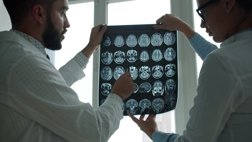
So, you’ve got a medical scan coming up, or maybe you’re just curious about how these incredible machines work. It’s easy to feel a bit in the dark when someone mentions an MRI, CT scan, or ultrasound. Think of this as your friendly guide to demystifying those whirring, beeping boxes that peek inside you.
At their core, medical scans are like highly sophisticated detective tools for doctors. They help visualize what’s happening inside your body without needing to actually go in there surgically. Each type of scan uses slightly different physics and technology to achieve this, but the goal is always the same: to get a clear picture so your doctor can figure out what’s going on and how best to help you.
X-rays: The Oldest Kid on the Block
When you think of medical imaging, X-rays are likely the first thing that comes to mind. They’ve been around for over a century, and for good reason – they’re useful for a whole range of things, from checking for broken bones to spotting pneumonia.
How X-rays Actually Work
It all starts with radiation. Don’t worry, it’s a very small dose, carefully controlled. An X-ray machine shoots a beam of these rays through your body. Think of it like shining a flashlight through different materials.
- Density Matters: Different tissues in your body have different densities. Bones are dense, so they absorb a lot of the X-ray. Soft tissues like muscles and organs are less dense and let more X-rays pass through. Air, like in your lungs, is very un-dense and lets almost all the X-rays through.
- Creating the Image: The X-rays that pass through your body then hit a detector (either film or a digital sensor). Where more X-rays hit, the image becomes darker. Where fewer X-rays make it through (like through bone), the image becomes lighter. This contrast is what allows us to see the different structures inside you.
What X-rays Are Good For
- Bones: This is their classic use. Detecting fractures, dislocations, and seeing any abnormalities in bone structure.
- Lungs: X-rays are excellent for spotting issues like pneumonia, fluid in the lungs, or masses.
- Digestive System (with contrast): Sometimes, you’ll be asked to swallow a special liquid (like barium). This coats your digestive tract, making it more visible on an X-ray and helping doctors see things like ulcers or blockages.
Limitations of X-rays
While versatile, X-rays aren’t great for seeing very soft tissues in detail. You won’t get a clear picture of your brain or muscles with a standard X-ray.
Medical scans, such as MRI and CT scans, utilize advanced technology to create detailed images of the body’s internal structures, aiding in diagnosis and treatment planning. These imaging techniques rely on principles of physics and biology, allowing healthcare professionals to visualize organs, tissues, and even blood flow without invasive procedures. For a deeper understanding of how these medical scans function and their significance in modern medicine, you can read a related article at this link.
CT Scans: Taking X-rays to the Next Level
CT stands forComputed Tomography. Imagine taking a whole bunch of X-ray images of a thin slice of your body from different angles and then using a computer to combine them into a detailed 3D picture. That’s essentially what a CT scan does.
The CT Scan Process
You’ll lie down on a table that slides into a large, circular machine. This machine houses an X-ray tube that rotates around you, taking hundreds of thin “slices” of your body.
- Rotational Imaging: As the X-ray tube circles, it sends out beams that are picked up by detectors on the opposite side. This happens very quickly, often in seconds.
- Computer Reconstruction: The real magic happens in the computer. It takes all these slice images and stitches them together, creating cross-sectional views. It’s like being able to look at your body as if you’ve sliced it up like a loaf of bread, but digitally.
- Contrast Agents: Just like with X-rays, sometimes a contrast dye is injected or swallowed. This further enhances the visibility of blood vessels and certain organs.
What CT Scans Excel At
- Trauma: CT scans are incredibly fast and great for quickly identifying injuries in complex areas like the head, chest, and abdomen, especially for internal bleeding or damage to organs.
- Cancer Detection and Staging: They can locate tumors, see how large they are, and whether they’ve spread to nearby lymph nodes or organs.
- Blood Vessels: With contrast, CT angiography can visualize blockages or abnormalities in arteries and veins.
- Complex Fractures: For intricate bone breaks, CT provides a more detailed view than a standard X-ray.
Types of CT Scans
- Head CT: Essential for diagnosing strokes, head injuries, and brain tumors.
- Chest CT: Used for lung diseases, heart conditions, and detecting masses.
- Abdominal CT: Helps identify problems with organs like the liver, kidneys, pancreas, and spleen.
MRIs: Leveraging Magnetism for Detail
MRI stands for Magnetic Resonance Imaging. This is where things get a bit different. Instead of X-rays, MRIs use powerful magnets and radio waves. This makes them particularly good at showing soft tissues in great detail.
The Science Behind MRI
Think of your body’s water molecules as tiny magnets. When you’re inside an MRI machine, a very strong magnetic field aligns these tiny magnets.
- Radio Wave Pulses: Then, short bursts of radio waves are sent through your body. These pulses knock the aligned water molecules out of position.
- Signal Detection: When the radio waves are turned off, the water molecules snap back into alignment with the main magnetic field, releasing their own radio signals.
- Computer Interpretation: Different tissues release slightly different signals based on their water content and how quickly they return to alignment. A computer processes these signals and creates incredibly detailed images.
Why MRIs Are So Good for Soft Tissues
- Brain and Spinal Cord: MRIs provide exceptional views of these areas, making them invaluable for diagnosing conditions like multiple sclerosis, herniated discs, and brain tumors.
- Joints and Muscles: They can clearly show ligaments, tendons, and muscles, which is crucial for diagnosing sports injuries or other musculoskeletal problems.
- Internal Organs: MRIs offer detailed images of organs like the liver, prostate, uterus, and breasts.
What to Expect During an MRI
MRI machines can be quite noisy, with loud banging and humming sounds. You’ll likely be given earplugs or headphones. Since it’s a strong magnetic field, you’ll be asked about any metal implants or jewelry you have.
Contrast in MRI
Like CT scans, MRIs can also use a contrast agent (often gadolinium-based) to highlight certain structures, particularly blood vessels and areas of inflammation.
Ultrasounds: Sounding Out Your Insides
Ultrasounds, also known as sonography, are perhaps the most familiar type of medical scan after X-rays. If you’ve ever seen a baby scan, you’ve seen an ultrasound in action. They use sound waves, not radiation!
The Magic of Sound Waves
Ultrasound works on a simple principle: sending sound waves into the body and listening for the echoes.
- The Transducer: A handheld device called a transducer is placed on your skin (usually with some gel to ensure good contact). This device both emits the sound waves and receives the echoes.
- Echoes as Information: The sound waves travel into your body and bounce off different tissues and organs. The transducer listens for these returning echoes.
- Building the Picture: The time it takes for the echoes to return and their strength are used by the machine to create an image. Dense structures, like bone or fluid, will create different echoes than softer tissues.
When Ultrasound Shines
- Pregnancy: This is its most famous application, allowing doctors to monitor fetal development.
- Abdominal Organs: It’s excellent for viewing the gallbladder, liver, kidneys, pancreas, and spleen, especially for detecting gallstones or cysts.
- Pelvic Organs: Commonly used to examine the uterus, ovaries, and bladder.
- Blood Flow: Doppler ultrasound can detect the speed and direction of blood flow in vessels, helping to identify blockages or abnormal flow.
- Soft Tissues: It can visualize superficial structures like the thyroid gland or lumps in the breast.
Advantages of Ultrasound
- No Radiation: This is a huge plus, making it safe for pregnant women and children.
- Real-time Imaging: You can see organs moving and blood flowing live on the screen.
- Portability: Many ultrasound machines are relatively portable, allowing for bedside scans.
Medical scans play a crucial role in diagnosing and monitoring various health conditions, utilizing advanced imaging technologies to visualize the internal structures of the body. For a deeper understanding of the principles behind these scans, you can explore a related article that delves into the intricacies of how these technologies function. This resource provides valuable insights into the different types of scans and their applications in modern medicine. To read more, visit this informative article that explains the fascinating world of medical imaging.
Nuclear Medicine Scans: Tracking Function, Not Just Structure
Nuclear medicine scans are a bit different from the others we’ve discussed so far. Instead of just looking at the physical structure of your organs, these scans focus on how well they’re working.
How Radioactive Tracers Work
These scans involve introducing a small amount of a radioactive substance, called a radiotracer or radionuclide, into your body.
- Targeting Specific Organs: Radiotracers are designed to be absorbed by specific organs or tissues. For example, a tracer might be chosen because it’s taken up by bone cells or by cancer cells.
- Emitting Gamma Rays: As the tracer breaks down over time, it emits tiny particles called gamma rays.
- Detection by a Scanner: A special camera (like a gamma camera or PET scanner) detects these gamma rays.
- Mapping Activity: The scanner maps where the radiation is concentrated, showing areas of high or low activity. This can reveal how an organ is functioning or where abnormal cells might be growing.
Common Types and Uses
- Bone Scans: Used to detect bone cancer, infections, or fractures that might not be visible on a standard X-ray.
- Thyroid Scans: Assess the function of the thyroid gland.
- Heart Scans (e.g., MPI – Myocardial Perfusion Imaging): Check blood flow to the heart muscle, particularly useful after a heart attack or for diagnosing coronary artery disease.
- PET Scans (Positron Emission Tomography): Often used in oncology to find cancer, see how it’s responding to treatment, and detect its spread. PET scans can also be used for neurological conditions like Alzheimer’s disease.
What Makes Them Unique
The key here is that nuclear medicine scans highlight biological activity. A structural scan might show a lump, but a nuclear scan might tell you if that lump is actively growing or metabolizing, which can be crucial for diagnosis and treatment.
Putting It All Together: Choosing the Right Scan
Your doctor will choose the best type of scan for your specific situation. There’s no single “best” scan; each has its strengths and weaknesses.
- For broken bones or pneumonia, an X-ray is often the first and best choice.
- For suspected internal bleeding after an accident or complex fractures, a CT scan offers speed and detail.
- To look closely at soft tissues like the brain, joints, or spinal cord, an MRI is usually preferred.
- To monitor a pregnancy, check for gallstones, or assess blood flow, an ultrasound is ideal.
- To understand how an organ is functioning or to detect specific types of cancer, nuclear medicine scans are invaluable.
Sometimes, a combination of scans might be used to get the most complete picture. The technology behind these scans is constantly evolving, leading to even clearer images and more accurate diagnoses. So, the next time you hear about a medical scan, you’ll have a better idea of the incredible science and technology at play, all working to help keep you healthy.

Why Modern Medicine Can’t Store Tomorrow
FAQs

1. What are medical scans used for?
Medical scans are used to create detailed images of the inside of the body. They are commonly used to diagnose and monitor various medical conditions, such as cancer, heart disease, and bone fractures.
2. How do X-rays work in medical scans?
X-rays work by passing a small amount of radiation through the body. Dense tissues, such as bones, absorb more radiation and appear white on the resulting image, while less dense tissues, such as muscles and organs, appear as shades of gray.
3. What is the difference between CT scans and MRI scans?
CT scans use X-rays to create cross-sectional images of the body, while MRI scans use a magnetic field and radio waves to produce detailed images of the body’s internal structures. CT scans are better for imaging bone and dense tissues, while MRI scans are better for soft tissues and organs.
4. Are medical scans safe?
While medical scans involve a small amount of radiation, the benefits of accurate diagnosis and treatment often outweigh the risks. However, it is important to discuss any concerns with a healthcare provider and to follow recommended safety guidelines.
5. How should patients prepare for a medical scan?
Patients may be asked to fast or avoid certain foods and drinks before a scan, depending on the type of scan being performed. It is important to follow any instructions provided by the healthcare provider and to inform them of any allergies or medical conditions.