So, you’ve heard about SPECT imaging and are wondering what exactly it is and how it pulls off its magic? Essentially, SPECT, which stands for Single-Photon Emission Computed Tomography, is a type of nuclear medicine scan that uses a radioactive tracer to show how blood flows or how organs are functioning. Think of it as a sophisticated internal mapping system that uses a tiny bit of safe radioactivity to reveal what’s happening deep inside your body, in 3D. It’s not about seeing structures like an MRI or CT scan; instead, it’s focused on revealing the activity of your cells and organs.
At its core, SPECT imaging is all about visualizing biological processes. Unlike regular X-rays that show solid structures, SPECT looks at function. We inject a small amount of a radioactive substance, called a radiotracer, into your body, usually intravenously. This radiotracer is designed to accumulate in specific organs or tissues that the doctor wants to examine. Once it’s in place, the tracer emits gamma rays, a type of photon. The SPECT scanner then detects these gamma rays from multiple angles, and a computer reconstructs this information into detailed 3D images. These images show where the radiotracer has gone and how concentrated it is, giving doctors a window into how active or healthy a particular area of your body is.
Injecting the Tracer: More Than Just a Shot
The radiotracer is the star of the show in SPECT. It’s not just any radioactive material; it’s carefully chosen for its properties.
Choosing the Right Tool for the Job
There are many different types of radiotracers, and the one used depends entirely on what the doctor is trying to see. For example, if they’re looking at blood flow to the heart, they might use a tracer like Technetium-99m sestamibi. If they’re investigating brain activity or looking for signs of Parkinson’s disease, they might opt for something else entirely, like Ioflupane I-123. The key is that each tracer has a “homing instinct” for specific cells or metabolic processes.
The Safety Factor: It’s Not Scary Radioactive
A common concern is radioactivity. It’s understandable! But it’s important to know that the amounts of radioactive material used in SPECT scans are very small, and they are considered safe for diagnostic purposes. The radioactivity decays quickly, meaning it loses its potency over a short period. For most SPECT tracers, the radiation dose is comparable to or even less than what you might receive from natural background radiation over a few months. Doctors and nuclear medicine technologists are trained to ensure the safe administration and handling of these materials.
Detecting the Signals: The SPECT Scanner at Work
Once the radiotracer has done its job and started emitting gamma rays, the SPECT scanner gets to work. It’s a rather impressive piece of equipment designed to capture these faint signals.
The Gamma Camera: The Scanner’s Eye
The core of a SPECT scanner is one or more gamma cameras. These sensitive detectors are equipped with special crystals (often sodium iodide) that emit light when struck by a gamma ray. This light is then amplified and converted into an electrical signal. The camera’s movement around the patient is crucial.
Rotating for a Full Picture
The SPECT scanner rotates around the patient, taking “pictures” from hundreds of different angles. Imagine shining a flashlight from every direction onto an object to paint a complete 3D portrait; it’s a similar principle. Each gamma camera captures the distribution of the radiotracer at a specific point in time. The more angles the scanner captures, the more accurate and detailed the resulting 3D image will be.
Spect imaging, or Single Photon Emission Computed Tomography, is a powerful imaging technique that allows for the visualization of functional processes in the body, particularly in the fields of cardiology and oncology. For a deeper understanding of how spect imaging works, including its applications and advancements, you can refer to this related article: Understanding Spect Imaging. This resource provides valuable insights into the technology behind spect imaging and its significance in modern medical diagnostics.
How the 3D Image is Built: From Dots to Depth
Capturing all those gamma ray signals is only half the battle. The real cleverness comes in how a computer takes that raw data and transforms it into a useful 3D image.
The Computer’s Reconstruction Act
The electrical signals generated by the gamma cameras are sent to a powerful computer. This is where the magic of tomography happens.
Back-Projection and Algorithms: The Nitty-Gritty
The computer uses complex mathematical algorithms to reconstruct the 3D image. One common technique is called filtered back-projection. Essentially, the computer “projects” the detected gamma rays back along the paths they traveled from the patient. By doing this from all the different angles, it can pinpoint where the radioactivity is concentrated within the body. Think of it like solving a puzzle where you have many overlapping clues, and the computer figures out the most likely arrangement to explain all the clues.
Slice by Slice: Building the Volume
The result of this reconstruction is a series of cross-sectional images, or “slices,” of the body. These slices, viewed from different orientations (like from the front, side, or top), are then stacked together to create a full 3D representation of the radiotracer’s distribution. This allows doctors to see precisely where the tracer has gone and in what concentration.
What SPECT Imaging Can Show Us

Because SPECT imaging focuses on function and blood flow, it’s incredibly useful for diagnosing and monitoring a wide range of conditions, especially those affecting organs with high metabolic activity.
Brainpower: Unlocking Neurological Mysteries
The brain utilizes a tremendous amount of energy, making it a prime candidate for SPECT imaging. It can provide vital information about how well blood is flowing to different parts of the brain and how active brain cells are.
Dementia and Alzheimer’s: Early Detection Clues
SPECT scans can help identify patterns of reduced blood flow or metabolic activity in the brain that are characteristic of certain forms of dementia, including Alzheimer’s disease. This can sometimes aid in earlier diagnosis and treatment planning.
Seizure Disorders: Pinpointing the Source
For individuals with epilepsy, SPECT can be particularly useful. By performing the scan during a seizure and then again when the person is seizure-free, doctors can compare the images. Areas with reduced blood flow when a person is seizure-free but increased blood flow during a seizure can help pinpoint the origin of the seizures, which is crucial for surgical planning.
Movement Disorders: Parkinson’s and Beyond
SPECT tracers that bind to dopamine transporters in the brain can be used to help diagnose and differentiate between various movement disorders, such as Parkinson’s disease and essential tremor. A reduced signal may indicate a loss of dopamine-producing cells, a hallmark of Parkinson’s.
Heart Health: A Look into the Engine Room
Your heart is a muscular organ that needs a constant supply of oxygenated blood to function. SPECT imaging is a valuable tool for assessing cardiac health.
Blood Flow During Stress: Stress Tests Made Visible
A common cardiac SPECT study involves injecting a radiotracer at rest and then again after the patient has undergone some form of stress, like exercise on a treadmill or medication to simulate exertion. By comparing the images, doctors can see if there are any areas of the heart muscle that don’t receive adequate blood flow during stress, which can indicate blockages in the coronary arteries.
Assessing Heart Damage: After a Heart Attack
SPECT can help determine the extent of damage to the heart muscle following a heart attack. It can show which areas of the heart are still viable (have good blood flow and function) and which areas have been permanently damaged.
Other Applications: Far-Reaching Insights
SPECT isn’t just for the brain and heart. It has applications in various other areas of medicine.
Bone Scans: Detecting Hidden Fractures and Infections
While plain X-rays are the first step for bone issues, SPECT bone scans can detect problems that X-rays might miss. This includes hairline fractures, stress fractures, infections in the bone (osteomyelitis), and certain types of bone cancer. The radiotracer accumulates in areas of increased metabolic activity, which often occurs with bone repair or inflammation.
Kidney Function: Assessing Drainage and Blood Flow
SPECT can be used to evaluate how well your kidneys are filtering blood and how well urine is draining from them. This can be important for diagnosing kidney stones, blockages, or assessing the function of transplanted kidneys.
Thyroid and Parathyroid Health: Balancing Hormones
SPECT imaging can also be used to assess the function of the thyroid and parathyroid glands, which are crucial for regulating metabolism and calcium levels in the body.
The SPECT Procedure: What to Expect

If you’re scheduled for a SPECT scan, it’s helpful to know what the process generally involves. It’s usually a straightforward and painless experience.
Before the Scan: Prep Work
Typically, there’s not a lot of preparation required for a standard SPECT scan.
Hydration and Medication: Simple Steps
You might be asked to drink plenty of fluids before your appointment, especially if you’re having a scan of your kidneys or urinary tract. It’s also important to let your doctor know about any medications you’re taking, as some might interfere with the scan or need to be adjusted. Usually, you can eat and drink normally beforehand, but always check with your doctor or the imaging center.
Understanding the Tracer Administration
The most significant part of the “prep” is the injection of the radiotracer. This is usually done via a small IV line inserted into a vein in your arm. You might feel a slight prick as the needle goes in, but the injection itself is generally painless.
During the Scan: Lies Back and Let It Scan
Once the radiotracer has been administered, there’s usually a waiting period. This allows the tracer to travel through your body and accumulate in the target organs. The duration of this waiting period varies widely depending on the radiotracer used and the organ being studied, ranging from as little as 15 minutes to several hours.
The Scanner’s Dance Around You
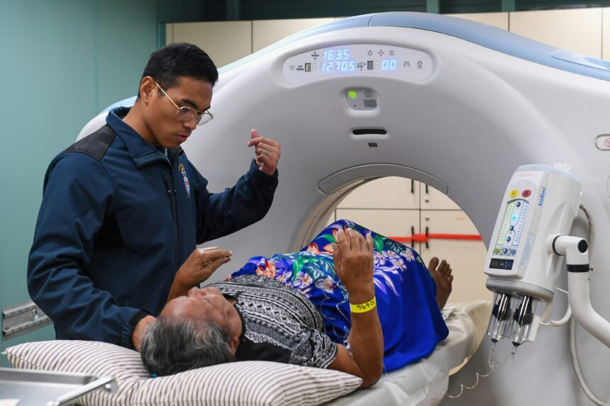
After the waiting period, you’ll be asked to lie down on a comfortable table, which will then slide into the center of the SPECT scanner. The scanner itself might look like a large donut or have rotating arms. You’ll need to remain as still as possible during the scan, as movement can blur the images. The scanner will slowly rotate around you, and you won’t feel anything during this process – no pain, no noise other than perhaps gentle whirring. The scan time itself can vary, but often ranges from 20 minutes to an hour.
After the Scan: Back to Normal
Once the scan is complete, you can usually resume your normal activities without any restrictions.
The Tracer Fades Away
As mentioned, the radiotracer quickly loses its radioactivity. Any remaining tracer will naturally pass out of your body over time, typically through urine. You’ll be advised to drink plenty of fluids to help flush it out.
Getting the Results: The Doctor’s Interpretation
The images are then reviewed by a radiologist or nuclear medicine physician who specializes in interpreting these scans. They will write a report detailing their findings, which will be sent to your referring doctor, who will then discuss the results with you.
Spect imaging, or Single Photon Emission Computed Tomography, is a sophisticated imaging technique that allows for the visualization of functional processes in the body by detecting gamma rays emitted from radiotracers. This method is particularly valuable in the fields of cardiology and oncology, as it provides insights into blood flow and metabolic activity. For a deeper understanding of the principles and applications of spect imaging, you can explore this informative article on the subject. By delving into the intricacies of how this technology works, you can appreciate its significance in modern medical diagnostics. To read more, visit this article.
SPECT vs. Other Imaging Techniques: Standing Out from the Crowd
| Component | Description |
|---|---|
| Radioactive tracer | A small amount of radioactive material is injected into the body, which emits gamma rays. |
| Gamma camera | Detects the gamma rays emitted by the radioactive tracer and creates images based on their distribution in the body. |
| Data processing | The collected gamma ray data is processed by a computer to create 3D images of the internal body structures. |
| Functional information | SPECT imaging provides information about blood flow, metabolism, and neurotransmitter activity in the body. |
SPECT imaging is a valuable tool, but it’s not the only game in town when it comes to medical imaging. Understanding how it differs from other common techniques can help clarify its unique role.
Structure vs. Function: The Core Difference
This is the most fundamental distinction.
CT and MRI: The Anatomical Architects
Computed Tomography (CT) and Magnetic Resonance Imaging (MRI) are primarily structural imaging techniques. They excel at showing detailed anatomical structures of the body – bones, organs, soft tissues, and blood vessels. A CT scan uses X-rays to create cross-sectional images, while MRI uses magnetic fields and radio waves. These are your go-to scans when a doctor wants to see the “shape” and “size” of something, like a tumor, a fracture, or an organ’s size and position.
PET Scans: A Close Cousin
Positron Emission Tomography (PET) is another nuclear medicine technique, sharing similarities with SPECT. Both use radiotracers to assess metabolic activity. The key difference lies in the type of radiation emitted and how it’s detected. PET tracers emit positrons, which then interact with electrons in the body to produce two gamma rays traveling in opposite directions. This allows for more precise localization and often higher resolution than SPECT. PET scans are often used for detecting cancer, assessing heart conditions, and studying brain disorders. Sometimes, SPECT and PET are combined (SPECT/CT or PET/CT) to merge anatomical and functional information.
SPECT’s Niche: When Function is Key
SPECT’s strength lies in its ability to visualize functional processes and blood flow over time.
Blood Flow Dynamics: Seeing the Flow
For certain applications, like assessing how well blood is getting to specific areas of the heart or brain, SPECT can be extremely informative. It can show differences in perfusion (blood supply) under different conditions, such as rest vs. stress in cardiac imaging.
Receptor Binding: Targeting Specific Molecules
Some SPECT tracers are designed to bind to specific receptors on cells. This is particularly useful in neurology for understanding the distribution of certain neurotransmitter pathways, as seen in Parkinson’s disease diagnosis. While PET also does this, SPECT can sometimes be more cost-effective and readily available.
Accessibility and Cost: Practical Considerations
In many healthcare settings, SPECT scanners are more widely available and generally less expensive to operate than PET scanners. This can make SPECT the preferred choice for certain diagnostic needs when PET isn’t strictly necessary for the level of detail required.
In conclusion, SPECT imaging is a powerful and versatile diagnostic tool that provides invaluable insights into the functional aspects of your body. By employing carefully selected radiotracers and sophisticated imaging technology, it allows doctors to see what’s really going on at a cellular and metabolic level, helping to diagnose a wide range of conditions and guide effective treatment.

Why Modern Medicine Can’t Store Tomorrow
FAQs
What is SPECT imaging?
SPECT (Single Photon Emission Computed Tomography) imaging is a nuclear medicine imaging technique that uses radioactive tracers to create 3D images of the body’s internal organs and tissues. It is commonly used to diagnose and monitor conditions such as heart disease, brain disorders, and bone abnormalities.
How does SPECT imaging work?
During a SPECT scan, a small amount of radioactive tracer is injected into the patient’s bloodstream. The tracer emits gamma rays, which are detected by a gamma camera as it rotates around the body. The data collected is then processed by a computer to create detailed images of the area being studied.
What are the benefits of SPECT imaging?
SPECT imaging provides valuable information about the function and structure of organs and tissues, allowing for early detection and accurate diagnosis of various medical conditions. It is non-invasive and can help guide treatment plans and monitor the effectiveness of therapies.
Are there any risks associated with SPECT imaging?
The amount of radiation exposure from a SPECT scan is considered to be low and generally safe for most patients. However, pregnant women and individuals with certain medical conditions may need to consult with their healthcare provider before undergoing a SPECT scan.
How long does a SPECT imaging procedure take?
The duration of a SPECT scan can vary depending on the specific area being studied and the type of tracer used. In general, the entire procedure, including preparation and imaging, may take anywhere from 1 to 3 hours.