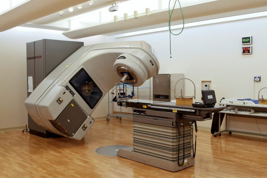
Nuclear medicine might sound a bit sci-fi, but it’s actually a super practical tool in healthcare. Think of it as using tiny, safe amounts of radioactive materials, called radiotracers, to see what’s happening inside your body at a molecular level. This gives doctors a really detailed picture of how your organs and tissues are functioning, which can be crucial for diagnosing and treating a whole range of conditions. It’s not about looking at static images like an X-ray; it’s about watching the body work.
Nuclear medicine isn’t just one single type of scan. It’s an umbrella term for several techniques that all involve radiotracers. The basic principle is pretty straightforward: the radiotracer is introduced into your body (usually by injection, but sometimes swallowed or inhaled), and it travels to specific organs or tissues. Because the radiotracer is radioactive, it emits tiny particles called gamma rays. Special cameras then detect these gamma rays and translate them into images or data that show how the radiotracer is being absorbed and distributed. This tells doctors about blood flow, organ function, and even if certain cells, like cancer cells, are unusually active.
The Role of Radiotracers
You might hear the terms “radioisotope” or “radiopharmaceutical.” These are essentially the same thing in this context. They are a combination of a radioactive atom and a molecule that has a biological purpose. For example, one common radiotracer uses a form of iodine that’s taken up by the thyroid gland. Another might use glucose tagged with a radioactive element, which is helpful because cancer cells tend to consume more glucose than healthy cells. The key is that these tracers are designed to be biologically active in a way that highlights what the doctor is looking for. They are given in very small, safe doses, and the radioactivity usually clears from your body fairly quickly.
Imaging Techniques: Different Ways to See
There are a few main ways that nuclear medicine images are created, each suited for different purposes:
SPECT Scans: A Detailed Slice
Single-Photon Emission Computed Tomography, or SPECT, is a bit like getting a 3D view of your body. Similar to CT scans, it takes multiple X-ray-like images from different angles and then uses a computer to reconstruct them into cross-sectional slices. This allows doctors to see the distribution of the radiotracer in much greater detail than a planar image, providing a clearer understanding of function in specific areas. It’s particularly useful for looking at blood flow in organs like the brain and heart, and for locating tumors.
PET Scans: Tracking Metabolism in Action
Positron Emission Tomography, or PET, is another powerful imaging technique that offers exceptional detail about metabolic activity. PET uses radiotracers that emit positrons. When a positron encounters an electron, they annihilate each other, producing two gamma rays that travel in opposite directions. The PET scanner detects these pairs of gamma rays simultaneously, allowing for very precise localization and quantification of the radiotracer. This is why PET scans are so effective at identifying areas of high metabolic activity, such as aggressive tumors or areas of inflammation.
Planar Imaging: The Basics
Sometimes, a simpler planar image is all that’s needed. Think of this as a straightforward picture showing where the radiotracer has gone and how much is there. While not as detailed as SPECT or PET, it’s a quick and effective way to assess the function of certain organs, like the thyroid or kidneys, and can be used to detect abnormalities.
For an insightful look into the intricacies of nuclear medicine and the technology that powers it, you can explore the article “Behind the Scenes of Nuclear Medicine” available at this link. This article delves into the processes and innovations that make nuclear medicine a vital tool in modern healthcare, highlighting the roles of various professionals and the cutting-edge equipment used in diagnostics and treatment.
Uncovering Disease: Diagnosis in Focus
The real power of nuclear medicine lies in its ability to detect diseases at their earliest stages, often before symptoms even appear, or to provide crucial information when other imaging methods aren’t conclusive.
Heart Health: A Vital Clue
One of the most common applications of nuclear medicine is in evaluating heart health.
Myocardial Perfusion Scans: Checking Blood Flow to the Heart
These scans are excellent for assessing how well blood is flowing to your heart muscle. You might be given a radiotracer and then have images taken while you’re at rest, and then again after some form of stress (either exercise or medication that makes your heart work harder). By comparing the images, doctors can identify areas of your heart muscle that aren’t receiving enough blood, which can indicate blockages in your coronary arteries, even if you’re not experiencing chest pain. This is a critical tool for diagnosing and managing coronary artery disease.
Evaluating Heart Valve Function
Nuclear medicine techniques can also be used to assess how well your heart valves are working, helping to diagnose conditions like valve regurgitation or stenosis.
Cancer Detection and Management: A Two-Pronged Approach
Nuclear medicine plays a significant role in the fight against cancer, both in finding it and in guiding treatment.
Pinpointing Tumors with FDG-PET
As mentioned earlier, many cancer cells have a higher metabolic rate and consume more glucose than normal cells. A common radiotracer used in PET scans is FDG (fluorodeoxyglucose), which is a type of sugar. When injected, FDG is taken up by cells that are actively using a lot of glucose. Areas that show high uptake of FDG are therefore suspicious for cancer. This can help doctors find the primary tumor, see if cancer has spread to other parts of the body (metastasis), and even determine if a tumor is cancerous or benign.
Assessing Treatment Effectiveness
After a cancer diagnosis, nuclear medicine can be invaluable in monitoring how well a treatment is working. If a tumor’s metabolic activity decreases after chemotherapy or radiation, it suggests the treatment is effective. Conversely, if the activity increases, it might mean the treatment needs to be adjusted. This “before and after” comparison is a powerful way to personalize cancer care.
Guiding Radiation Therapy
In some cases, nuclear medicine can pinpoint the exact location and extent of a tumor, which helps radiation oncologists plan the most precise radiation therapy treatments, minimizing damage to surrounding healthy tissues.
Bone Scans: More Than Just Fractures
Bone scans are another well-established use of nuclear medicine, providing a much more detailed view of bone health than standard X-rays.
Detecting Occult Fractures
Sometimes, a fracture can be very subtle and not clearly visible on an X-ray, especially hairline fractures or those in complex bone structures. A bone scan can detect these by showing areas of increased bone turnover, which is a sign of healing and often indicates a fracture. This is particularly useful in cases of suspected abuse or in athletes with stress fractures.
Identifying Infections and Inflammation
Bone scans can also highlight areas of infection (osteomyelitis) or inflammation in the bones, which might be difficult to diagnose otherwise. The radiotracer will accumulate in these areas as the body responds to the abnormal activity.
Locating Metastatic Cancer in Bones
Cancer that has spread to the bones (bone metastases) is a common and serious complication of several types of cancer. Bone scans are very sensitive in detecting these metastases, often picking them up before they cause pain or are visible on X-rays.
Thyroid and Kidney Function: Essential Insights
Nuclear medicine provides direct insights into the functioning of these vital organs.
Thyroid Scans: Understanding Hyper- and Hypothyroidism
A thyroid scan uses radioactive iodine to assess the size, shape, and function of the thyroid gland. It can help diagnose hyperthyroidism (overactive thyroid) and hypothyroidism (underactive thyroid), as well as identify nodules or goiters.
Kidney Scans: Evaluating Renal Function
Renal scintigraphy uses radiotracers to measure how well your kidneys are filtering waste from your blood and how quickly urine is draining from them. This can help diagnose kidney infections, blockages, or damage from conditions like hypertension or diabetes.
Beyond Diagnosis: Therapeutic Applications
While imaging is a major part of nuclear medicine, it also has a powerful therapeutic side, using radioactivity to treat diseases. This is often referred to as “theranostics” – a blend of therapeutics and diagnostics.
Targeted Cancer Therapy: Radioactive “Magic Bullets”
This is where nuclear medicine truly shines in treatment.
Iodine-131 for Thyroid Cancer
For certain types of thyroid cancer, radioactive iodine (I-131) is used as a treatment. After surgery to remove the thyroid, patients are given a dose of I-131, which is preferentially absorbed by any remaining thyroid cancer cells, destroying them. This is a highly effective and targeted form of treatment.
Radionuclide Therapy for Other Cancers
Other radioactive isotopes are attached to molecules that specifically target cancer cells, acting like tiny radioactive “bullets” that deliver radiation directly to the tumor while sparing healthy tissues. Examples include treatments for metastatic prostate cancer and certain neuroendocrine tumors.
Pain Management: Relieving Bone Metastasis Aches
For patients with painful bone metastases, certain radiopharmaceuticals can be administered intravenously. These compounds are taken up by areas of increased bone turnover, including the sites of cancer spread, delivering radiation to the bone and helping to reduce pain.
Treating Overactive Thyroid Disease
In cases of hyperthyroidism, a controlled dose of radioactive iodine (I-131) can be given. The radioactive iodine is absorbed by the overactive thyroid cells, gradually destroying them and bringing thyroid hormone levels back to normal. This is an effective alternative to surgery or daily medication for many patients.
The Safety of Nuclear Medicine: Understanding the Risks

It’s natural to be concerned about radiation, but it’s important to understand that nuclear medicine procedures are designed with safety as a top priority.
Doses and Decay: Minimizing Exposure
The amount of radioactivity used in diagnostic nuclear medicine scans is extremely small. In fact, the radiation dose from many SPECT and PET scans is comparable to what you might receive from a few days of natural background radiation. The radiotracers used are also chosen for their short half-lives, meaning they decay and lose their radioactivity relatively quickly. For example, some radiotracers have a half-life of only a few hours, meaning half of their radioactivity is gone in that time.
Regulatory Oversight: Strict Guidelines
Nuclear medicine facilities and professionals are highly regulated by government agencies. These regulations ensure that doses are kept as low as reasonably achievable (ALARA principle), imaging procedures are appropriate for the patient’s condition, and waste is handled safely. Technologists are highly trained in radiation safety and handling protocols.
Pregnancy and Breastfeeding: Special Considerations
For pregnant women, nuclear medicine procedures are generally avoided unless absolutely necessary and if the potential benefit outweighs the risk to the fetus. For breastfeeding mothers, specific protocols are in place, often involving temporarily stopping breastfeeding for a period after the scan to allow the radiotracer to clear from the body. Your doctor will discuss these considerations thoroughly with you.
Nuclear medicine plays a crucial role in diagnosing and treating various medical conditions, and understanding the intricacies of this field can be fascinating. For those interested in exploring the behind-the-scenes aspects of nuclear medicine, a related article can provide valuable insights. You can read more about the innovative techniques and technologies used in this field by visiting this article, which delves into the processes that make nuclear medicine an essential component of modern healthcare.
What to Expect During a Nuclear Medicine Procedure
| Aspect | Details |
|---|---|
| Radioisotopes | Used in nuclear medicine for imaging and treatment |
| Production | Radioisotopes are produced in nuclear reactors or cyclotrons |
| Imaging Techniques | Includes SPECT, PET, and gamma camera imaging |
| Medical Professionals | Specialized nuclear medicine physicians and technologists |
| Regulations | Strict regulations for handling and disposal of radioactive materials |
While the specifics can vary depending on the type of scan, here’s a general idea of what you might experience.
Before the Scan: Preparation is Key
The most common preparation involves the administration of the radiotracer. This is usually done via an intravenous injection into a vein in your arm. You’ll then need to wait for a period, known as the uptake or distribution time, ranging from a few minutes to a few hours, to allow the radiotracer to travel through your body and reach the target area. During this waiting period, you might be asked to relax and avoid strenuous activity. Depending on the type of scan, you might be asked to drink water to help clear the radiotracer from your bladder or to empty your bladder before the scan.
During the Scan: Staying Still is Important
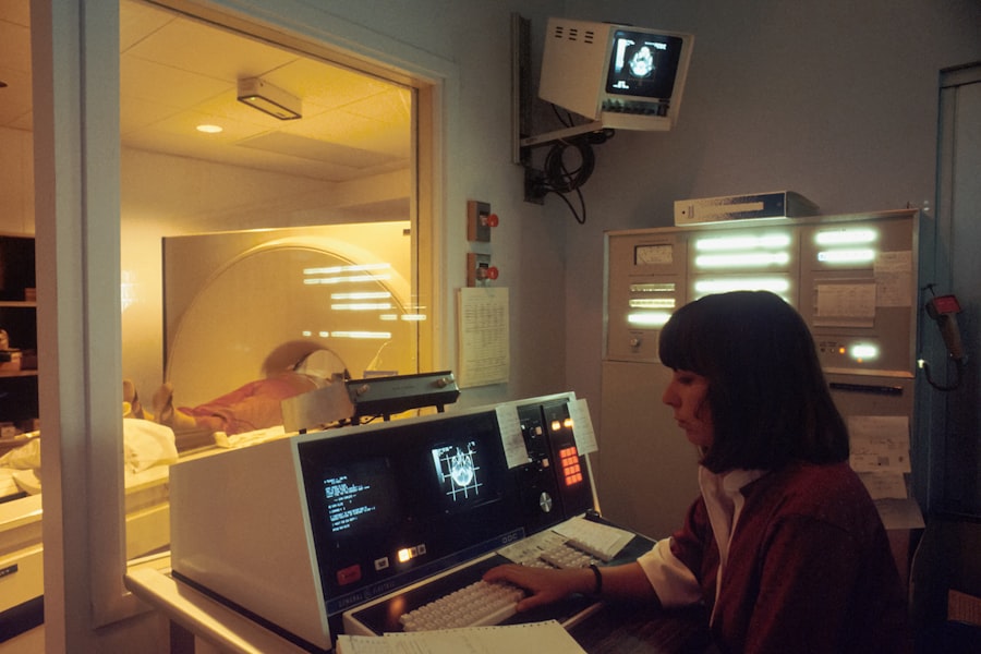
Once the radiotracer has had time to distribute, you’ll be positioned on a special imaging table. The cameras used in nuclear medicine, like SPECT and PET scanners, are typically large, doughnut-shaped machines that you’ll lie inside of. It’s crucial to remain as still as possible during the scan to ensure the images are clear and accurate. You’ll be able to communicate with the technologist throughout the procedure. The scan itself can last anywhere from 20 minutes to over an hour, depending on the complexity of the imaging required.
After the Scan: Resuming Normal Activities
For most diagnostic nuclear medicine scans, you can resume your normal activities immediately after the procedure. The radiotracer will continue to be eliminated from your body through urine and feces, and you’ll usually be advised to drink plenty of fluids for the first few hours to help with this process. The small amount of radiation from the tracer dissipates very quickly. If you’ve undergone radionuclide therapy, the recovery and any necessary precautions will be explained in detail by your medical team.
In essence, nuclear medicine offers a remarkable window into the inner workings of the body, providing invaluable information for both diagnosis and treatment. It’s a sophisticated field that continues to evolve, offering hope and better outcomes for patients facing a wide array of health challenges.

Why Modern Medicine Can’t Store Tomorrow
FAQs
What is nuclear medicine?
Nuclear medicine is a medical specialty that uses small amounts of radioactive materials, or radiopharmaceuticals, to diagnose and treat a variety of diseases and conditions within the body.
How does nuclear medicine work?
In nuclear medicine, a patient is given a small amount of a radioactive substance, which then travels to the specific area of the body being examined. A special camera detects the radiation emitted from the radiopharmaceuticals and creates images that help diagnose and treat the patient’s condition.
What are the common uses of nuclear medicine?
Nuclear medicine is commonly used to diagnose and treat conditions such as cancer, heart disease, thyroid disorders, and bone abnormalities. It can also be used to evaluate organ function and detect certain types of infections.
What are the benefits of nuclear medicine?
Nuclear medicine allows for early detection and treatment of diseases and conditions, often before symptoms occur. It also provides valuable information about the function of organs and tissues, which can help guide treatment decisions.
What are the potential risks of nuclear medicine?
The amount of radiation used in nuclear medicine procedures is considered safe for patients and healthcare providers. However, there is a small risk of allergic reactions to the radiopharmaceuticals, as well as a very small risk of radiation exposure. Patients should discuss any concerns with their healthcare provider before undergoing a nuclear medicine procedure.