So, you’re curious about nuclear medicine? It sounds a bit sci-fi, doesn’t it? But at its core, it’s really about using safe, tiny amounts of radioactive materials to help doctors see what’s going on inside your body in a way that regular X-rays or MRIs can’t. Think of it as a super-sensitive insider’s view, helping to diagnose diseases earlier and more accurately, and even treat certain conditions.
Let’s break down how this whole “nuclear medicine” thing works. It’s not about blowing anything up; it’s more about a delicate dance between science and your body.
Small Stuff, Big Impact: Radiopharmaceuticals
The key players in nuclear medicine are called radiopharmaceuticals. Don’t let the name scare you – they’re essentially specialized drugs containing a tiny amount of a radioactive atom, called a radionuclide. This radionuclide is attached to a molecule that has a specific job, like finding cancer cells or showing how organs are functioning.
- Targeted Delivery: The molecule is designed to go to a particular place in your body. For example, some might be attracted to bone, others to specific types of tumors, or areas with increased blood flow.
- The “Tag”: The radioactive part is like a tiny, invisible “tag” that can be detected by special cameras. It emits a form of radiation called gamma rays.
Seeing the Unseen: Detection Methods
Once the radiopharmaceutical is in your body, it does its job, and then the magic of detection happens.
- Gamma Cameras are Key: The most common imaging technique uses a gamma camera (sometimes called a scintillation camera). This is a sophisticated device that captures the gamma rays emitted by the radiopharmaceutical.
- Building a Picture: As the camera scans your body, it records the gamma rays, and a computer uses this information to create detailed images. These images show where the radiopharmaceutical has accumulated, which tells doctors a lot about what’s happening at a cellular level.
- More Than Just Pictures: Some nuclear medicine scans, like PET scans, provide even more information. They don’t just show where something is; they can also show how active it is, giving a sense of biological processes in action.
Nuclear medicine is a fascinating field that utilizes radioactive materials for diagnosis and treatment of various medical conditions. For a deeper understanding of the principles and applications of nuclear medicine, you can explore a related article that discusses its mechanisms and advancements in the field. To read more, visit this article.
Why Radiopharmaceuticals are Safe (Surprisingly!)
One of the first questions people usually have is about safety. “Radioactive? Isn’t that dangerous?” It’s a valid concern, and the answer is nuanced but ultimately reassuring.
Tiny Doses, Big Difference
The amount of radioactive material used in nuclear medicine imaging is incredibly small. It’s just enough to be detected by the cameras, but not enough to cause harm.
- Short Half-Life Heroes: Many of the radionuclides used have very short half-lives. This means they quickly decay and lose their radioactivity. For example, some might have a half-life of only a few hours. By the time you leave the clinic, much of the radioactivity will have already gone.
- Processed Naturally: Your body also processes and eliminates the radiopharmaceutical, usually through urine or stool, further reducing any residual exposure.
Weighing the Risks and Benefits
Like any medical procedure, there’s always a very small risk associated with radiation exposure. However, the benefits of a nuclear medicine scan – getting a diagnosis, understanding the extent of a disease, or guiding treatment – almost always far outweigh these minimal risks.
- Radiation Equivalence: The amount of radiation you receive from a typical nuclear medicine scan is often comparable to, or even less than, what you might receive from natural background radiation over a few days or weeks.
The Diagnostic Arsenal: What Can it Find?
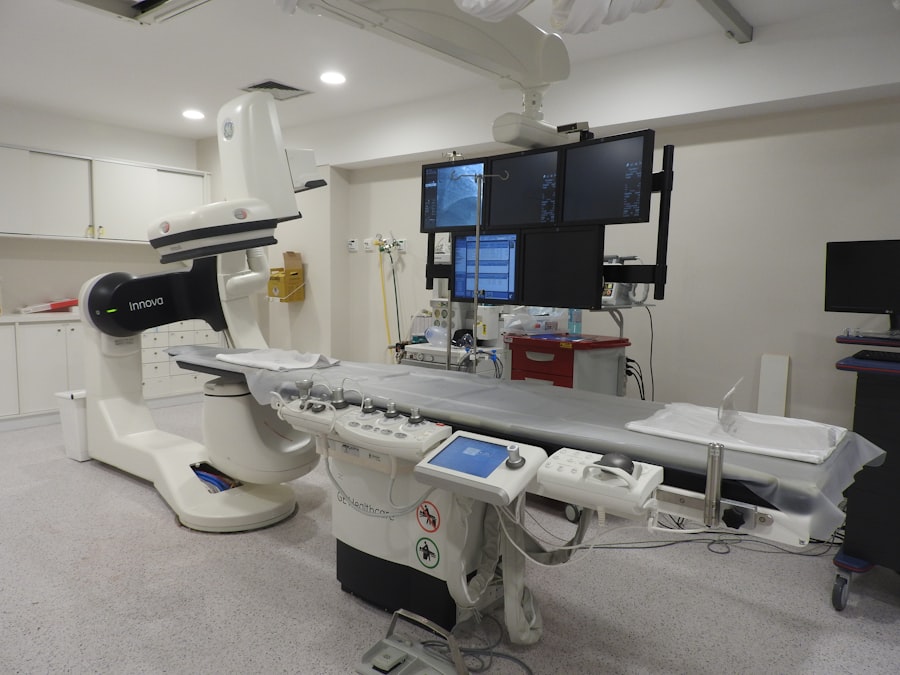
Nuclear medicine isn’t just one tool; it’s a versatile set of techniques used to diagnose a wide range of conditions, often when other methods fall short.
Illuminating Cancer’s Footprint
One of the most significant applications of nuclear medicine is in cancer diagnosis and management.
- Spotting the Small Stuff: Radiopharmaceuticals can often detect cancerous cells very early, sometimes even before a tumor is visible on other imaging scans. This is because cancer cells often have a higher metabolic rate and “take up” certain tracers more readily.
- Staging and Monitoring: Nuclear medicine scans are vital for determining the stage of cancer (how far it has spread) and for monitoring how well a treatment is working. If cancer cells are still active after treatment, it can show up on a scan.
Beyond Cancer: A Spectrum of Uses
While cancer detection is a major area, nuclear medicine plays a crucial role in diagnosing and managing many other diseases.
- Heart Health: It can show how well blood is flowing to your heart muscle, helping to diagnose and assess the severity of coronary artery disease and to evaluate the impact of heart attacks.
- Brain Power: It can help identify problems in the brain, likeAlzheimer’s disease, Parkinson’s disease, and epilepsy, by showing areas of altered brain activity or blood flow.
- Bone Issues: Bone scans are excellent for detecting stress fractures, infections, and bone cancer, especially when symptoms are vague.
- Kidney Function: Nuclear medicine can assess how well your kidneys are working and identify blockages or other issues.
- Thyroid Troubles: It’s a cornerstone in diagnosing and treating thyroid conditions, including hyperthyroidism and thyroid cancer.
Therapeutic Power: Healing with Radiation
Nuclear medicine isn’t just about looking inside; it can also be used to treat diseases. This is known as radionuclide therapy.
Targeted Radiation Therapy
In radionuclide therapy, a larger dose of a radioactive substance is used to directly target and destroy diseased cells.
- Radioactive Isotopes for Treatment: Similar to diagnostic procedures, a radioactive isotope is attached to a molecule that is designed to accumulate in specific areas. However, the radionuclide used here is chosen for its ability to deliver a therapeutic dose of radiation.
- Killing Diseased Cells: The radiation emitted by the radionuclide damages the DNA of the targeted cells, leading to their destruction. This is particularly effective for certain types of cancer where the diseased cells have a specific marker that the radiopharmaceutical can bind to.
Common Therapeutic Applications
- Thyroid Cancer and Hyperthyroidism: Radioactive iodine ($^{131}$I therapy) has been a remarkably successful treatment for both overactive thyroid glands (hyperthyroidism) and thyroid cancer for decades. The thyroid gland naturally takes up iodine, so the radioactive iodine selectively targets thyroid cells.
- Prostate Cancer: Radiopharmaceutical therapy is increasingly used for advanced prostate cancer. Certain radiopharmaceuticals bind to prostate-specific membrane antigen (PSMA), a protein that is often overexpressed on prostate cancer cells.
- Pain Management: For patients with bone metastases that cause significant pain, certain radiopharmaceuticals can be injected to deliver radiation to these painful lesions, providing relief.
The Experience of Treatment
Radionuclide therapy is typically administered orally (as a pill or liquid) or intravenously. Patients may need to be hospitalized for a short period to ensure radiation safely dissipates before returning home, depending on the type and dose of the radionuclide used.
Nuclear medicine is a fascinating field that utilizes radioactive materials to diagnose and treat various medical conditions. By introducing small amounts of radioactive substances into the body, healthcare professionals can obtain detailed images of organs and tissues, allowing for accurate assessments of health. For a deeper understanding of the principles and applications of nuclear medicine, you can explore this informative article on the subject. It delves into the techniques and technologies that make this medical specialty so effective in patient care. To read more, visit this article.
The Patient Experience: What to Expect
| Aspect | Description |
|---|---|
| Radioactive Tracers | Radioactive substances are injected into the body to visualize and diagnose diseases. |
| Gamma Cameras | Specialized cameras detect the gamma rays emitted by the radioactive tracers. |
| Images | The gamma camera creates images that show the distribution of the radioactive tracers in the body. |
| Diagnostics | Physicians use these images to diagnose and monitor various medical conditions. |
| Treatment | Nuclear medicine can also be used for therapeutic purposes, such as treating certain types of cancer. |
If you’re scheduled for a nuclear medicine procedure, it’s natural to wonder what the actual experience is like. It’s generally straightforward and quite different from what you might imagine from the “nuclear” part of the name.
Before the Scan: Preparation is Key
Your doctor or the nuclear medicine department will give you specific instructions, but here are some common preparatory steps.
- Dietary Restrictions: Sometimes, you might be asked to avoid certain foods or drinks for a period before the scan. For example, if you’re having a thyroid scan, you might be asked to avoid seafood or dairy for a few days.
- Medication Review: It’s crucial to inform your healthcare provider about all medications you are taking, as some can interfere with the scan.
- Hydration: Often, being well-hydrated before the scan is recommended.
During the Scan: Relax and Be Still
The actual imaging part is usually quite simple and doesn’t involve much discomfort.
- Administration of the Radiopharmaceutical: This is typically done by injection, though sometimes it can be taken orally or inhaled. You might feel a slight prick from the injection, but that’s usually it.
- Waiting Period: After the radiopharmaceutical is administered, there’s often a waiting period. This allows the substance to travel through your body and reach its target. The length of this wait varies widely depending on the type of scan, from about 15 minutes to a few hours. During this time, you can usually relax, read, or do quiet activities.
- The Imaging: You’ll lie down on a comfortable table, and the gamma camera or PET scanner will move around you. It’s important to stay as still as possible during the scan to ensure clear images. The cameras don’t touch you, and the process is painless. The scanning time itself can range from 20 minutes to over an hour.
After the Scan: Getting Back to Normal
Once the scan is complete, you’re usually free to go about your usual activities.
- No Special Precautions: For most diagnostic scans, there are no special precautions you need to take. You can eat, drink, and interact with others as normal. The small amount of radioactivity is harmless to those around you.
- Increased Fluid Intake: You might be encouraged to drink plenty of fluids to help your body eliminate the radiopharmaceutical.
- Results: Your doctor will discuss the results of your scan with you, usually within a few days.
The Future of Nuclear Medicine: Always Evolving
Nuclear medicine is a field that’s constantly innovating. Researchers are always looking for new ways to use this powerful technology for even better diagnosis and treatment.
Sharper Imaging, Better Targets
- New Radiotracers: Scientists are developing new radiopharmaceuticals that can bind to even more specific targets within the body. This means more precise identification of diseases and a better understanding of their behavior.
- Advanced Imaging Technology: Newer generations of PET and SPECT scanners are offering higher resolution and faster scan times, leading to more detailed and informative images.
Personalized Medicine and Beyond
- Theranostics: A major area of growth is “theranostics,” which combines diagnostic imaging with targeted therapy. This allows doctors to identify diseased cells with a diagnostic tracer and then, if they have the right characteristics, treat them with a therapeutic radiopharmaceutical. This is a highly personalized approach to medicine.
- Earlier Detection and Prevention: The ultimate goal is to use nuclear medicine to detect diseases at the earliest possible stage, when they are most treatable, and potentially even to identify individuals at high risk for developing certain conditions.
Nuclear medicine, with its ability to peer into the body at a functional level and its growing therapeutic applications, is a vital and evolving part of modern healthcare. It offers a unique window into our health, helping doctors make better decisions and leading to improved outcomes for patients.

Why Modern Medicine Can’t Store Tomorrow
FAQs
What is nuclear medicine?
Nuclear medicine is a medical specialty that uses small amounts of radioactive materials, or radiopharmaceuticals, to diagnose and treat a variety of diseases and conditions within the body.
How does nuclear medicine work?
In nuclear medicine, a patient is given a small amount of a radioactive substance, which is then detected by a special camera that creates images of the inside of the body. These images can show how organs and tissues are functioning, rather than just how they look.
What are some common uses of nuclear medicine?
Nuclear medicine is commonly used to diagnose and treat conditions such as cancer, heart disease, and certain types of infections. It can also be used to evaluate organ function and to assess the effectiveness of treatments.
Is nuclear medicine safe?
The amount of radiation used in nuclear medicine procedures is considered safe for patients. The benefits of the information gained from these procedures usually outweigh the risks of radiation exposure.
What are the advantages of nuclear medicine over other imaging techniques?
Nuclear medicine can provide unique information about the function of organs and tissues, which is not always possible with other imaging techniques such as X-rays, CT scans, or MRI. It can also help doctors make more accurate diagnoses and develop more targeted treatment plans.